Chapter 5. Sensation and Perception
Hearing
Jessica Motherwell McFarlane
Approximate reading time: 20 minutes
Our auditory system converts pressure waves into meaningful sounds. This translates into our ability to hear the sounds of nature, to appreciate the beauty of music, and to communicate with one another through spoken language. This section will provide an overview of the basic anatomy and function of the auditory system. It will include a discussion of how sensory stimulus is translated into neural impulses, where in the brain that information is processed, how we perceive pitch, and how we know where sound is coming from.
Anatomy of the Auditory System
The ear can be separated into multiple sections. The outer ear includes the pinna, which is the visible part of the ear that extends from our heads, the auditory canal, and the tympanic membrane (also known as the eardrum). The middle ear contains three tiny bones known as the ossicles, which are named the malleus (or hammer), incus (or anvil), and the stapes (or stirrup). The inner ear contains the semicircular canals, which are involved in balance and movement (the vestibular sense), and the cochlea. The cochlea is a fluid-filled, snail-shaped structure that contains the sensory receptor cells (hair cells) of the auditory system. Within the cochlea lies the basilar membrane, a critical component that plays a key role in hearing. This membrane runs the entire length of the cochlea and vibrates in response to sound waves. The vibrations of the basilar membrane are crucial for the conversion of sound waves into electrical signals that can be interpreted by the brain. Different frequencies of sound waves cause specific regions of the basilar membrane to vibrate, allowing for the perception of a wide range of sounds. This precise mechanism enables the auditory system to distinguish between various pitches and tones (Figure SP.12).
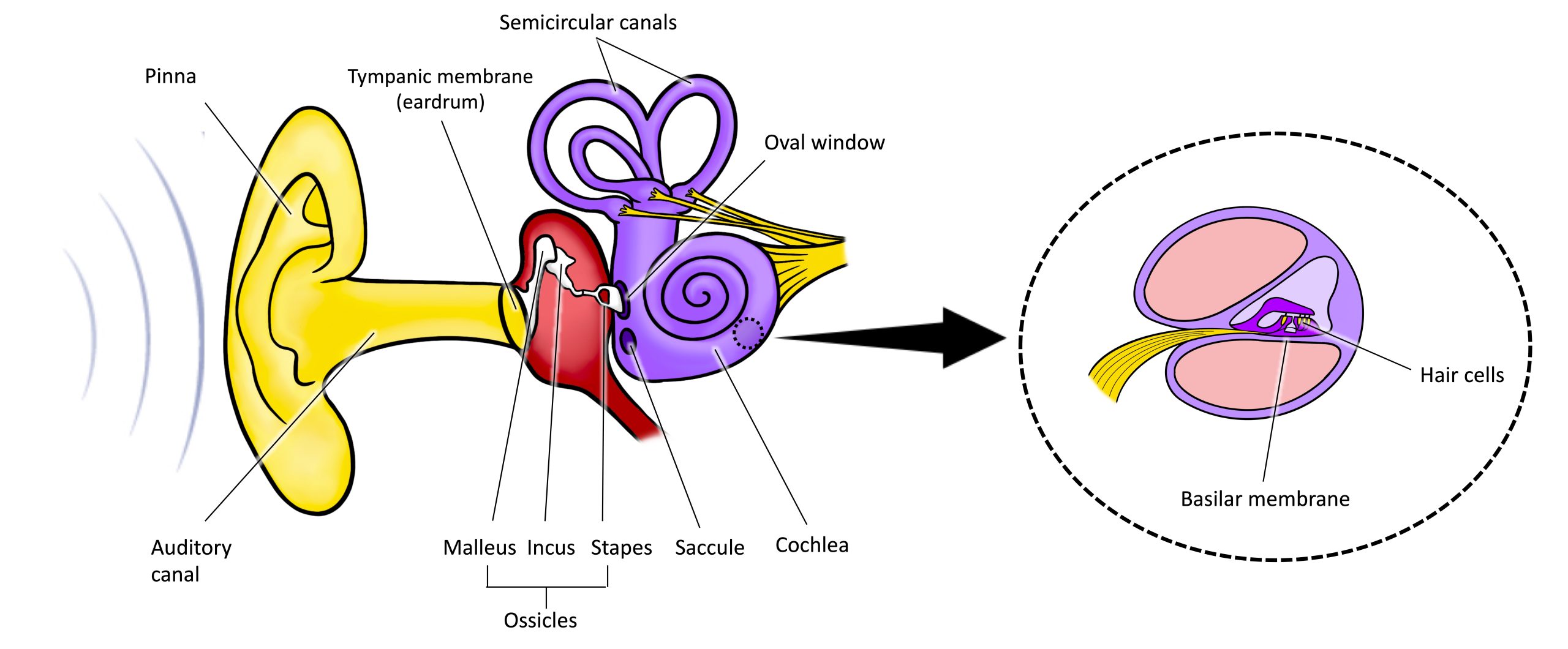
Sound waves travel along the auditory canal and strike the tympanic membrane, causing it to vibrate. This vibration results in movement of the three ossicles (malleus, incus, stapes). As the ossicles move, the stapes presses into a thin membrane of the cochlea known as the oval window. As the stapes presses into the oval window, the fluid inside the cochlea begins to move, which in turn stimulates hair cells, which are auditory receptor cells of the inner ear embedded in the basilar membrane. The basilar membrane is a thin strip of tissue within the cochlea.
The activation of hair cells is a mechanical process: the stimulation of the hair cell ultimately leads to activation of the cell. As hair cells become activated, they generate neural impulses that travel along the auditory nerve to the brain. Auditory information is shuttled to the inferior colliculus, the medial geniculate nucleus of the thalamus, and finally to the auditory cortex in the temporal lobe of the brain for processing. Like the visual system, there is also evidence suggesting that information about auditory recognition and localization is processed in parallel streams (Rauschecker & Tian, 2000; Renier et al., 2009).
Watch this video: Hearing & Balance: Crash Course Anatomy & Physiology #17 (10.5 minutes)
“Hearing & Balance: Crash Course Anatomy & Physiology #17” video by CrashCourse is licensed under the Standard YouTube licence.
Hearing Loss
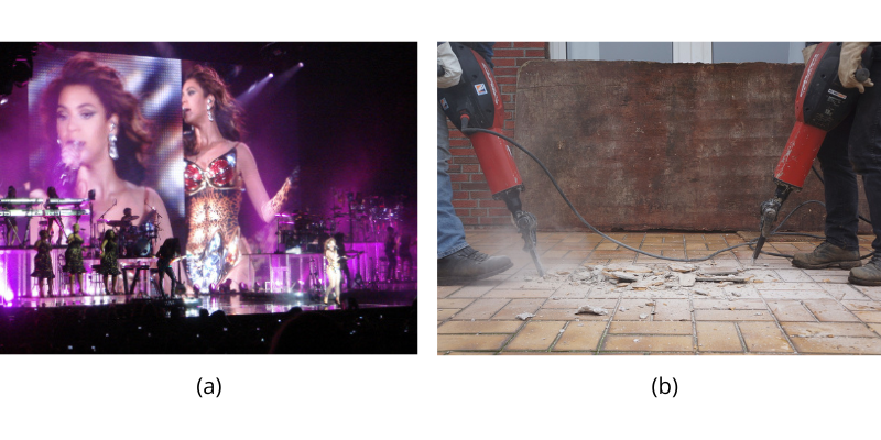
Deafness is the partial or complete inability to hear. Some people are born without hearing, which is known as congenital deafness. Other people have conductive hearing loss, which is due to a problem delivering sound energy to the cochlea. Causes for conductive hearing loss include blockage of the ear canal, a hole in the tympanic membrane, problems with the ossicles, or fluid in the space between the eardrum and cochlea. Another group of people experience sensorineural hearing loss, which is the most common form of hearing loss. Sensorineural hearing loss can be caused by many factors, such as ageing, head or acoustic trauma, infections and diseases (such as measles or mumps), medications, environmental effects such as noise exposure (noise-induced hearing loss, as shown in Figure SP.13), tumours, and toxins (such as those found in certain solvents and metals).
Given the mechanical nature by which the sound wave stimulus is transmitted from the eardrum through the ossicles to the oval window of the cochlea, some degree of hearing loss is inevitable. With conductive hearing loss, hearing deficits are associated with a lack of vibration of the eardrum and/or movement of the ossicles. These symptoms can be alleviated by using devices like hearing aids that amplify incoming sound waves to make vibration of the eardrum and movement of the ossicles more likely to occur.
When the hearing problem is associated with a failure to transmit neural signals from the cochlea to the brain, it is called sensorineural hearing loss. One disease that results in sensorineural hearing loss is Ménière’s disease. Although not well understood, Ménière’s disease results in a degeneration of inner ear structures that can lead to hearing loss, tinnitus (constant ringing or buzzing), vertigo (a sense of spinning), and an increase in pressure within the inner ear (Semaan & Megerian, 2011). This kind of loss cannot be treated with hearing aids, but some individuals might be candidates for a cochlear implant as a treatment option.
Deaf Culture
In Canada, the United States, and other places around the world, Deaf people have their own language, schools, and customs. This is called Deaf Culture. In Canada, there are two official sign languages: American Sign Language (ASL) and Quebec Sign Language, la langue des signes quebecoise (LSQ). ASL has no verbal component and is based entirely on visual signs and gestures. The primary mode of communication is signing. One of the values of Deaf Culture is to continue traditions like using sign language rather than teaching Deaf children to try to speak, read lips, or have cochlear implant surgery.
Deafness, traditionally viewed as a handicap leading to discrimination, has been redefined through the idea of “deaf gain” introduced by Bauman and Murray (2014). Recent research affirms that sign language and Deaf Culture are essential for the social, emotional, and cognitive development of Deaf individuals (Bogdanova, 2021; Humphries, et al., 2023; Tomaszewski, et al., 2019).
Deaf Gain
Deaf gain is a concept that reframes deafness, highlighting the unique advantages and perspectives it offers rather than viewing it solely as a loss of hearing. It suggests that the absence of sound can enhance other senses and modes of communication, leading to enriched visual and tactile sensitivities and advanced skills in visual-spatial tasks and sign language. Deaf gain also celebrates the cultural and social contributions of the Deaf community, emphasising its rich art, history, and values. This perspective encourages a broader understanding of sensory perception, informed by individual experiences and beliefs, and underscores the importance of diversity and accessible communication in enriching society.
Sign language is much more than a mere literal translation of spoken languages; it is the practical and social centre of Deaf Culture. With unique syntax and semantics, it’s a distinct language in its own right (Padden & Humphries, 2005). There are hundreds of sign languages worldwide, each with its own structure, grammar, and vocabulary. Studies reveal that sign language not only supports cognitive development and social skills but can also enhance spoken language development for native signing children with cochlear implants (Humphries et al., 2012).
Cochlear implants are electronic devices that consist of a microphone, a speech processor, and an electrode array. The device receives incoming sound information and directly stimulates the auditory nerve to transmit information to the brain. Cochlear implants have been met with a mix of acceptance and resistance within the Deaf community. While some parents of deaf children may embrace this technology as a tool for facilitating communication in a predominantly hearing world, others within the Deaf community may reject such interventions, viewing them as incompatible with their Deaf identity and culture (Sparrow, 2005). This divide reflects broader debates about deafness, identity, and the importance of preserving Deaf culture and sign language (Kusters et al., 2017; Leigh, 2010).
The rights of Deaf people are supported by the United Nations Convention on the Rights of Persons with Disabilities (CRPD), which acknowledges sign language as equal to spoken language (United Nations, 2006). Engaging with the Deaf community means understanding and respecting their culture and language. This involves practicing appropriate etiquette, such as maintaining eye contact during conversation, ensuring adequate lighting for clear sign visibility, and getting attention in a respectful manner, like a gentle tap on the shoulder or a wave (Stewart & Akamatsu, 2002). Learning basic sign language helps in communicating effectively with Deaf individuals and fosters a sense of understanding and appreciation for their unique culture.
Image Attributions
Figure SP.12. Figure SAP.18 created by Molly Wells Art as found in Introduction to Psychology & Neuroscience (2nd Edition) is licensed under a CC BY 4.0 License.
Figure SP.13. Figure 5.9 as found in Psychology 2e by OpenStax is licensed under a CC BY 4.0 License and contains modifications of the following works: (a) Beyonce I am… Tour 2009 Live in Berlin by GillyBerlin is licensed under a CC BY 2.0 License. (b) Image by c_badeja is used under the Pixabay Content License.
To calculate this time, we used a reading speed of 150 words per minute and then added extra time to account for images and videos. This is just to give you a rough idea of the length of the chapter section. How long it will take you to engage with this chapter will vary greatly depending on all sorts of things (the complexity of the content, your ability to focus, etc).
