Chapter 4. Biological Basis of Behaviour
Cells of the Nervous System
Tareq Yousef
Approximate reading time: 66 minutes
Learning Objectives
By the end of this section, you will be able to:
- Appreciate some of the history associated with studying the brain
- Be aware of the basic parts of a neuron and familiarise yourself with the vocabulary of the nervous system
- Consider how neurons communicate with each other
- Recognise how drugs act as agonists or antagonists for a given neurotransmitter system
The History of Studying Brains is Old!
Present day psychologists and neuroscientists interested in the workings of the brain have access to advanced technology (covered in-depth later in this chapter) to attempt to answer questions about the brain’s workings. But, their efforts did not reach this modern state without guidance from scientists before them. Less credit is given to some of the earlier neuroanatomists (those who study the anatomy of the nervous system) like Hasan Ibn al-Haytham, also known as Alhazen (born 965). A Persian scientist, al-Haytham is credited with what is possibly the world’s first known illustration of the nervous system.

Interested in vision, al-Haytham argued against hundreds of years of theories by Euclid and Ptolemy, renowned Greek and Alexandrian scholars respectively, that suggested vision was due to our eyes emitting light (Lindberg, 1967). By theorising that vision requires light to actually enter our eyes, al-Haytham got closer to what modern scientists have confirmed.
The illustration of the beginnings of the visual system credited to al-Haytham depicts broadly the components of the eye (which you will become familiar with later in this chapter) but also the eye’s physical connection with something behind it… the processing of the brain! In fact, you will learn that the light-sensitive tissue at the back of the eye, the retina, is just an outgrowth from our brain that extends during early development. This image and the writing of al-Haytham are the only historical evidence, an indication that curiosity about the brain is much older than the invention of the microscope.
Modern Understanding of the Human Brain
Psychologists striving to understand the human mind may study the nervous system. Learning how the body’s cells and organs function can help us understand the biological basis of human psychology. The nervous system is composed of two basic cell types: glia and neurons. Glia (a single one is a glial cell) are traditionally thought to play a supportive role to neurons, both physically and metabolically (i.e., supplying nutrients and maintaining their function). Glia provide scaffolding on which the nervous system is built, help neurons line up closely with each other to allow neuronal communication, provide insulation to neurons, transport nutrients and waste products, and mediate immune responses.
For years, researchers believed that there were many more glia than neurons; however, more recent work from Suzanna Herculano-Houzel’s laboratory has called this long-standing assumption into question and has provided important evidence that there may be a nearly 1:1 ratio of glia to neurons. This is important because it suggests that human brains are more similar to other primate brains than previously thought (Azevedo et al, 2009; Hercaulano-Houzel, 2012; Herculano-Houzel, 2009). Neurons, on the other hand, serve as interconnected information processors that are essential for all of the tasks of the nervous system. This means that these cells can act as different parts of a machine (the entire nervous system) to carry out a function (like moving your leg, or retrieving a memory). This section briefly describes the structure and function of neurons.
Neuron Structure
Neurons are the central building blocks of the nervous system, 100 billion strong at birth. Like all cells, neurons consist of several different parts, each serving a specialised function (Figure BB.2). A neuron’s outer surface is made up of a semipermeable membrane. The semipermeable nature of the membrane allows smaller molecules and molecules without an electrical charge to pass through it, while stopping larger or highly charged molecules.
, axon, and terminal buttons. A myelin sheath covers part of the neuron.")
The nucleus of the neuron is located in the soma, or cell body. The soma has branching extensions known as dendrites. The neuron is a small information processor, and dendrites serve as input sites where signals are received from other neurons. These signals are transmitted electrically across the soma and down a major extension from the soma known as the axon, which ends at multiple terminal buttons (also commonly referred to as boutons–the French word for buttons). The terminal buttons contain synaptic vesicles that house neurotransmitters, the chemical messengers of the nervous system.
Axons range in length from a fraction of an inch to several feet. In some axons, glia form a fatty substance known as the myelin sheath, which coats the axon and acts as an insulator, increasing the speed at which the signal travels. The myelin sheath is not continuous and there are small gaps that occur down the length of the axon. These gaps in the myelin sheath are known as the Nodes of Ranvier. The myelin sheath is crucial for the normal operation of the neurons within the nervous system; the loss of the insulation it provides can be detrimental to normal function.
To understand how this works, let’s consider an example. Phenylketonuria (fen-ul-key-toe-NU-ree-uh), also called PKU, causes a reduction in myelin and abnormalities in white matter cortical and subcortical structures (regions of the brain below the surface). This disorder is associated with a variety of issues including severe cognitive deficits (i.e., difficulties with everyday functioning or learning), exaggerated reflexes, and seizures (Anderson & Leuzzi, 2010; Huttenlocher, 2000). Another disorder, multiple sclerosis (MS), an autoimmune disorder, involves a large-scale loss of the myelin sheath on axons throughout the nervous system. The resulting interference in the electrical signal prevents the quick transmission of information by neurons and can lead to a number of symptoms, such as dizziness, fatigue, loss of motor control, and sexual dysfunction. While some treatments may help to modify the course of the disease and manage certain symptoms, there is currently no known cure for multiple sclerosis.
Watch this video: Tricky Topics: Neuronal Structure (17.5 minutes)
“Tricky Topics: Neuronal Structure” video by FirstYearPsych Dalhousie is licensed under the Standard YouTube licence.
Here is the Tricky Topics: Neuronal Structure transcript.
In typically functioning nervous systems, the neuronal signal moves rapidly down the axon to the terminal buttons, where synaptic vesicles release neurotransmitters into the synaptic cleft (Figure BB.3). The synaptic cleft is a very small space between two neurons and is an important site where communication between neurons occurs. Once neurotransmitters are released into the synaptic cleft, they travel across it and bind with corresponding receptors on the dendrite of an adjacent neuron. Receptors, proteins on the cell surface where neurotransmitters attach, vary in shape, with different shapes matching different neurotransmitters.
How does a neurotransmitter know which receptor to bind to? The neurotransmitter and the receptor have what is referred to as a lock-and-key relationship — specific neurotransmitters fit specific receptors similar to how a key fits a lock. The neurotransmitter binds to any receptor that it fits.
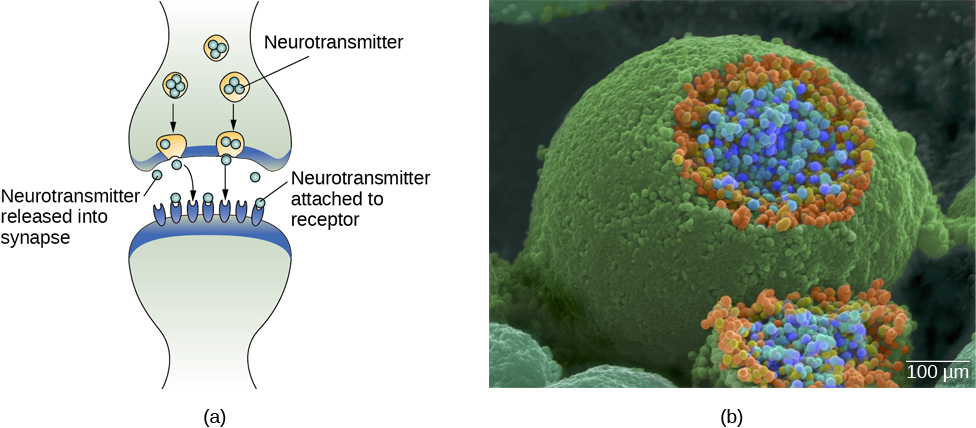
Figure BB.3. Neurotransmitter. The synaptic cleft is the space between the terminal button of one neuron and the dendrite of another neuron. (b) In this pseudo-coloured image from a scanning electron microscope, a terminal button (green) has been opened to reveal the synaptic vesicles (orange and blue) inside. Each vesicle contains about 10,000 neurotransmitter molecules.Watch this video: Tricky Topics: Synaptic Transmission (9 minutes)
“Tricky Topics: Synaptic Transmission” video by FirstYearPsych Dalhousie is licensed under the Standard YouTube licence.
Here is the Tricky Topics: Synaptic Transmission transcript.
Neuronal Communication
Now that we have learned about the basic structures of the neuron and the role that these structures play in neuronal communication, let’s take a closer look at the signal itself — how it moves through the neuron and then jumps to the next neuron, where the process is repeated.
We start our journey at the neuronal membrane. The neuron exists in a fluid environment; it is surrounded by fluid outside the cell, called extracellular fluid, and contains fluid inside the cell, known as intracellular fluid or cytoplasm. The neuronal membrane acts as a barrier, keeping these two fluids separate. This role is crucial because the electrical signal passing through the neuron relies on the electrical difference between the intra- and extracellular fluids. This charge difference, called the membrane potential, supplies the energy for the signal.
The electrical charge of the fluids is caused by charged molecules (ions) dissolved in the fluid. The semipermeable nature of the neuronal membrane somewhat restricts the movement of these charged molecules, and, as a result, some of the charged particles tend to become more concentrated either inside or outside the cell.
Between signals, the neuron membrane’s potential is held in a state of readiness, called the resting potential. The typical resting potential of a neuron is -70 millivolts (mV). Like a rubber band stretched out and waiting to spring into action, ions line up on either side of the cell membrane, ready to rush across the membrane when the neuron goes active and the membrane opens its gates (i.e., a sodium-potassium pump that allows movement of ions across the membrane). Ions in high-concentration areas are ready to move to low-concentration areas in a process called osmosis (you may have come across the term before), and positive ions are ready to move to areas with a negative charge. This is a fundamental process, where negatively charged particles are attracted to positive charges, just like the interaction of two magnets.
In the resting state, sodium (Na+) is at higher concentrations outside the cell, so it will tend to move into the cell. Potassium (K+), on the other hand, is more concentrated inside the cell, and will tend to move out of the cell (Figure BB.4). In addition, the inside of the cell is slightly negatively charged compared to the outside. This difference in charge, known as the electrochemical gradient, acts on sodium, causing it to move into the cell.

From this resting potential state, the neuron receives a signal and its state changes abruptly (Figure BB.5). When a neuron receives signals at the dendrites — due to neurotransmitters from an adjacent neuron binding to its receptors — small pores, or gates, open on the neuronal membrane, allowing Na+ ions, propelled by both charge and concentration differences, to move into the cell. With this influx of positive ions, the internal charge of the cell becomes more positive. If that charge reaches a certain level, called the threshold of excitation, the neuron becomes active and the action potential begins.
Several additional pores open, leading to a massive influx of positively charged sodium ions creating a significant spike in the membrane potential, the peak of the action potential. At this peak, the sodium gates close, and the potassium gates open. Positively charged potassium ions start to exit the cell rapidly, initiating the process of repolarization. Initially, the cell becomes more negative than its resting potential, known as hyperpolarization, and then it stabilises, returning to its resting potential.

This positive spike represents the action potential: the electrical signal that typically moves from the cell body down the axon to the axon terminals. The electrical signal moves down the axon with the impulses jumping in a leapfrog fashion between the Nodes of Ranvier. The Nodes of Ranvier are natural gaps in the myelin sheath. At each point, some of the sodium ions that enter the cell diffuse to the next section of the axon, raising the charge past the threshold of excitation and triggering a new influx of sodium ions. The action potential moves all the way down the axon in this way until reaching the terminal buttons.
The action potential is an all-or-none phenomenon. In simple terms, this means that an incoming signal from another neuron is either sufficient or insufficient to reach the threshold of excitation. There is no in-between, and there is no turning off an action potential once it starts. Think of it like sending an email or a text message. You can think about sending it all you want, but the message is not sent until you hit the send button. Furthermore, once you send the message, there is no stopping it.
Since it’s an all-or-none response, the action potential is reproduced, or propagated, at its full strength along every point of the axon. Imagine it like a lit fuse on a firecracker — it doesn’t lose intensity as it travels down the axon. This all-or-none property explains why your brain perceives an injury to a distant body part, such as your toe, as equally painful as one to your nose (which is closer to your brain).
As noted earlier, when the action potential arrives at the terminal button, the synaptic vesicles release their neurotransmitters into the synaptic cleft. The neurotransmitters travel across the synapse and bind to receptors on the dendrites of the adjacent neuron, and the process repeats itself in the new neuron (assuming the signal is sufficiently strong to trigger an action potential). Once the signal is delivered, excess neurotransmitters in the synaptic cleft drift away, break down into inactive fragments (degradation), or are reabsorbed in a process known as reuptake.
Reuptake involves the neurotransmitter being pumped back into the neuron that released it, in order to clear the synapse (Figure BB.6). Clearing the synapse serves both to provide a clear “on” and “off” state between signals and to regulate the production of neurotransmitter (full synaptic vesicles signal that no additional neurotransmitters need to be produced). The synapse can also be cleared via degradation of the neurotransmitter, which typically involves an enzyme breaking the neurotransmitter down into its components, so that it can no longer interact with the receptors on the postsynaptic neuron.

Neuronal communication is often referred to as an electrochemical event. The movement of the action potential down the length of the axon is an electrical event, and movement of the neurotransmitter across the synaptic space represents the chemical portion of the process. However, there are some specialised connections between neurons that are entirely electrical. In such cases, the neurons are said to communicate via an electrical synapse. In these cases, two neurons physically connect to one another via gap junctions that allow the current from one cell to pass into the next. There are far fewer electrical synapses in the brain, but those that do exist are much faster than the chemical synapses that have been described above (Connors & Long, 2004).
Watch this video: Tricky Topics: Action Potentials (18 minutes)
“Tricky Topics: Action Potentials” video by FirstYearPsych Dalhousie is licensed under the Standard YouTube licence.
Here is the Tricky Topics: Action Potentials transcript.
Neurotransmitters and Drugs
There are several different types of neurotransmitters released by different neurons, and we can speak in broad terms about the kinds of functions associated with different neurotransmitters (Table BB.1). Much of what psychologists know about the functions of neurotransmitters comes from research on the effects of drugs in psychological disorders. Psychologists who take a biological perspective and focus on the physiological causes of behaviour assert that psychological disorders like depression and schizophrenia are associated with imbalances in one or more neurotransmitter systems. From this perspective, psychotropic medications can help improve the symptoms associated with these disorders. Psychotropic medications are drugs that treat psychiatric symptoms by restoring neurotransmitter balance.
| Neurotransmitter | Involved in | Potential effect on behaviour |
|---|---|---|
| Acetylcholine | Muscle activity, memory | Increased arousal, enhanced cognition |
| Beta-endorphin | Pain, pleasure | Decreased anxiety, decreased tension |
| Dopamine | Mood, sleep, learning | Increased pleasure, suppressed appetite |
| Gamma-aminobutyric acid (GABA) | Inhibitory control, sleep | Decreased anxiety, decreased tension |
| Glutamate | Memory, learning | Increased learning, enhanced memory |
| Norepinephrine | Heart function, intestinal function, alertness | Increased arousal, suppressed appetite |
| Serotonin | Mood, sleep | Modulated mood, suppressed appetite |
Psychoactive drugs can act as agonists or antagonists for a given neurotransmitter system. Agonists are chemicals that mimic a neurotransmitter at the receptor site. An antagonist, on the other hand, blocks or impedes the normal activity of a neurotransmitter at the receptor. Agonists and antagonists represent drugs that are prescribed to correct the specific neurotransmitter imbalances underlying a person’s condition. For example, Parkinson’s disease, a progressive nervous system disorder, is associated with low levels of dopamine. Therefore, a common treatment strategy for Parkinson’s disease involves using dopamine agonists, which mimic the effects of dopamine by binding to dopamine receptors.
Specific symptoms of schizophrenia are linked to increased dopamine neurotransmission. The medications used to treat these symptoms are called antipsychotics. They work as dopamine antagonists, which means they block dopamine’s effects by attaching to its receptors without activating them. As a result, they prevent dopamine released by one neuron from transmitting information to nearby neurons.
In contrast to agonists and antagonists, which both operate by binding to receptor sites, reuptake inhibitors prevent unused neurotransmitters from being transported back to the neuron. This allows neurotransmitters to remain active in the synaptic cleft for longer durations, increasing their effectiveness. Depression, which has been consistently linked with reduced serotonin levels, is commonly treated with selective serotonin reuptake inhibitors (SSRIs). By preventing reuptake, SSRIs strengthen the effect of serotonin, giving it more time to interact with serotonin receptors on dendrites. Common SSRIs include Prozac, Paxil, and Zoloft. The drug LSD is structurally very similar to serotonin, and it affects the same neurons and receptors as serotonin.
Psychotropic drugs are not instant solutions for people suffering from psychological disorders. Often, an individual must take a drug for several weeks before seeing improvement, and many psychoactive drugs have significant negative side effects. Furthermore, individuals vary dramatically in how they respond to drugs. To improve chances for success, it is not uncommon for people receiving pharmacotherapy (drugs for therapeutic purposes) to undergo psychological and/or behavioural therapies as well. Some research suggests that combining drug therapy with other forms of therapy tends to be more effective than any one treatment alone (for one such example, see March et al., 2007).
Image Attributions
Figure BB.1. Alhazen1652.png is in the public domain.
Figure BB.2 created by Molly Wells Art as found in Introduction to Psychology & Neuroscience (2nd Edition) is licensed under a CC BY 4.0 License.
Figure BB.3. Figure 3.9 as found in Psychology 2e by OpenStax is licensed under a CC BY 4.0 License and contains modifications of the following works: a) Neurotransmitter activity by OpenStax is licensed under a CC BY-NC-SA 4.0 license. b) modification of work by Tina Carvalho, NIH-NIGMS; scale-bar data from Matt Russell. Image is in the public domain.
Figure BB.4. Figure 3.10 as found in Psychology 2e by OpenStax is licensed under a CC BY 4.0 License.
Figure BB.5. Figure 3.11 as found in Psychology 2e by OpenStax is licensed under a CC BY 4.0 License.
Figure BB.6. Figure 3.12 as found in Psychology 2e by OpenStax is licensed under a CC BY 4.0 License.
To calculate this time, we used a reading speed of 150 words per minute and then added extra time to account for images and videos. This is just to give you a rough idea of the length of the chapter section. How long it will take you to engage with this chapter will vary greatly depending on all sorts of things (the complexity of the content, your ability to focus, etc).
nervous system cells that provides physical and metabolic support to neurones, including neuronal insulation and communication, and nutrient and waste transport
cells in the nervous system that act as interconnected information processors, which are essential for all of the tasks of the nervous system
cell membrane that allows smaller molecules or molecules without an electrical charge to pass through it, while stopping larger or highly charged molecules
cell body
branch-like extension of the soma that receives incoming signals from other neurones
major extension of the soma
axon terminal containing synaptic vesicles
storage site for neurotransmitters
chemical messenger of the nervous system
fatty substance that insulates axons
open spaces that are found in the myelin sheath that encases the axon
small gap between two neurones where communication occurs
protein on the cell surface where neurotransmitters attach
difference in charge across the neuronal membrane
the state of readiness of a neurone membrane’s potential between signals
level of charge in the membrane that causes the neurone to become active
electrical signal that moves down the neurone’s axon
level of charge in the membrane that causes the neurone to become active
neurotransmitter is pumped back into the neurone that released it
the process by which an enzyme breaks down neurotransmitters in the synaptic cleft into their components so that they can no longer interact with the receptors on the postsynaptic neurone.
view that psychological disorders like depression and schizophrenia are associated with imbalances in one or more neurotransmitter systems
drugs that treat psychiatric symptoms by restoring neurotransmitter balance
drug that mimics or strengthens the effects of a neurotransmitter
drug that blocks or impedes the normal activity of a given neurotransmitter
