1.3 Research Methods in Psycholinguistics
Psycholinguistics employs a number of ways understand language. These range from observational studies, speech error analysis to experiments and neuroimaging techniques. We also use computational models to simulate our theories about the language system. This section will explore some of the techniques employed by researchers. However, keep in mind that we are always developing new techniques to understand how language works.
Mental Chronometry
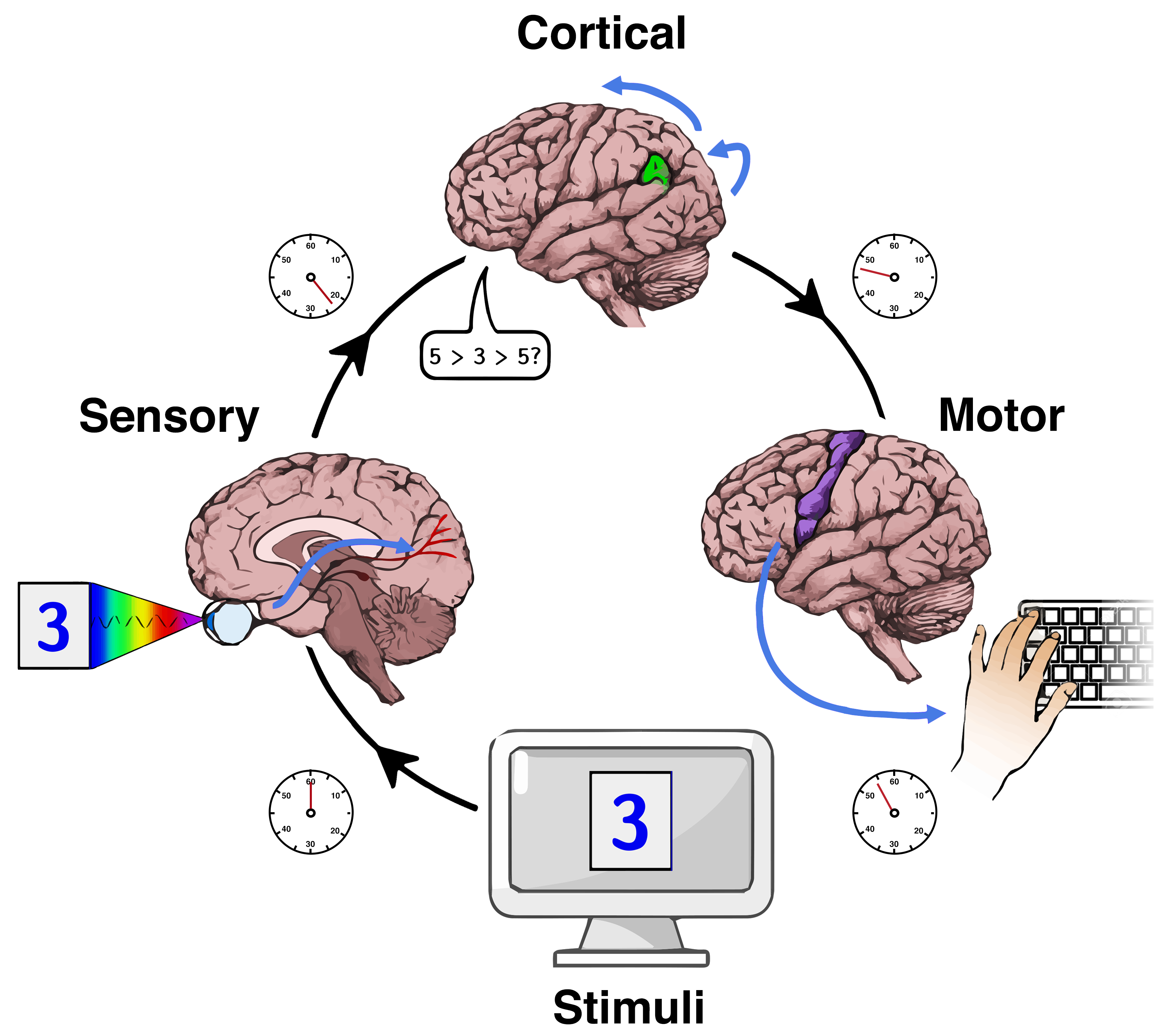
The study of reaction time on cognitive tasks is a common psychological paradigm in trying to infer the duration, sequence and content of cognition. As seen in Figure 1.5, reaction time (or RT) is measured as the time between the onset of a stimuli and the response by the participant. The mean and the variance of reaction times are considered useful indices on processing speed. The most common form of reaction time experiments are button presses. However, eye movements and voice onset (in repetition and reading tasks) can also be employed.
One of the most popular reaction time paradigms is called priming. Priming is used in almost all areas of psychology. The basic idea is that if two things share some cognitive or psychological attribute, they will either facilitate or interfere with each other. However, it they do not share such similarities, there will be no such effect. For example, it is easier to recognize the word DOG if you have already seen the word CAT. This can be a kind of sematic priming in that both words belong to the same semantic category (ANIMAL). Such an effect is known as facilitation while the interference of slowing down of such an effect if known as interference.

As seen in Figure 1.6, the reasoning behind priming effects can be modelled as a web of interconnected ideas or concepts in the mind. Concepts that are connected semantically (dogs and frogs are both animals) or phonologically (dog and bog end with similar sounds) are more likely to facilitate priming. In Figure 1.6, sematic connections are indicated with straight lines while phonological connects are indicated with dotted lines. The idea is that encountering a stimulus (by seeing or hearing it) will not only activate that concept in the mind but also partially activate connected concepts to some degree. As such, when any one of those connected concepts is presented next, they will be retrieved quicker because they have already been partially activated (or primed) by the previous activation.
Lesion Studies
As the brain is a vulnerable organ, it can be damaged by external or internal trauma. If blood flow and oxygen supply is constricted even for a few minutes to neurons they begin to die. These sites of damage are called lesions. Such trauma can be from accidents, strokes, brain surgery, or the ingestion of certain toxins. Examining these lesions and associating them with the behavioural limitations of such patients can provide valuable information about which regions are responsible for which behaviour. Cognitive Neuropsychology has contributed to psycholinguistics from the earliest times. Perhaps the earliest record of this is from case 20 in the Edwin Smith Papyrus. It is the report of a patient with a head injury which led to the following observation: “…He is speechless. An ailment not to be cured.” A clear case of speech loss due to brain injury. Centuries later, Broca and Wernicke continue with such observation and we will discuss them in Chapter 4. Cognitive neuropsychology attempts to relate brain-damaged behavioural deficits to models of normal processing. Shallice (1988) overserved that cognitive neuropsychology has made significant advances in associating neurological disorders to cognitive model, emphasized the importance of single case studies over group studies, and contributed to the exploration of impaired brain behaviour as a way towards understanding unimpaired behaviour. While traditional lesion studies were conducted by post-mortem examination and backtracking to analyse the behaviour of the patient while alive, modern neuroimaging techniques allow us to examine lesions in patients while they are alive and conduct behavioural analysis in real time.
Electroencephalography (EEG)
The advent of neuroimaging techniques has led to a flowering of new research in psycholinguistics. While traditional X-rays are not able to provide much detail on the brain, other technology such as the measurement of electrical activity in the brain have provided valuable data. Such techniques include electroencephalography or EEG which measures the brain’s electrical activity by detecting them from electrodes placed on the scalp. An amplifier can then amplify the millivoltage differences across the scalp and provide a continuous reading of brain activity.
Psychologists go even further and measure such electrical activity by tying them to specific events (such as the presentation of a stimulus). Such event-related potential or ERPs can have positive or negative polarities. These peaks in ERP readings are labelled according to their polarity (positive or negative) and the time difference from the stimuli onset (in milliseconds). Some common ERPs include N400 (detected 400ms after stimulus onset as a negative voltage) and P300 (detected 600ms after stimulus onset as a positive voltage). As EEG and ERP are measuring electrical activity, they detect changes in the brain almost instantly. We can say they have very good temporal resolution. However, as they are detecting this electrical potential from the scalp, the signals that are detected tend to be an averaged out one from multiple brain regions and neurons. Therefore, it is not always possible to pinpoint which brain region was actually involved in a particular EEG or ERP signal. In other words, these techniques have poor spatial resolution. Other techniques such as PET and MRI have been developed as a way to increase the spatial resolution of neuroimaging.
Positron Emission Tomography (PET)
PET (positron emission tomography) uses radioactive substances as tracers to produce images of brain activity. As the brain consumes a large amount of energy, injecting glucose into the body ensures that most of it ends up in brain regions that are active in a cognitive task. If the glucose contains isotopes that are radioactive, their emissions can be detected and transformed into images.
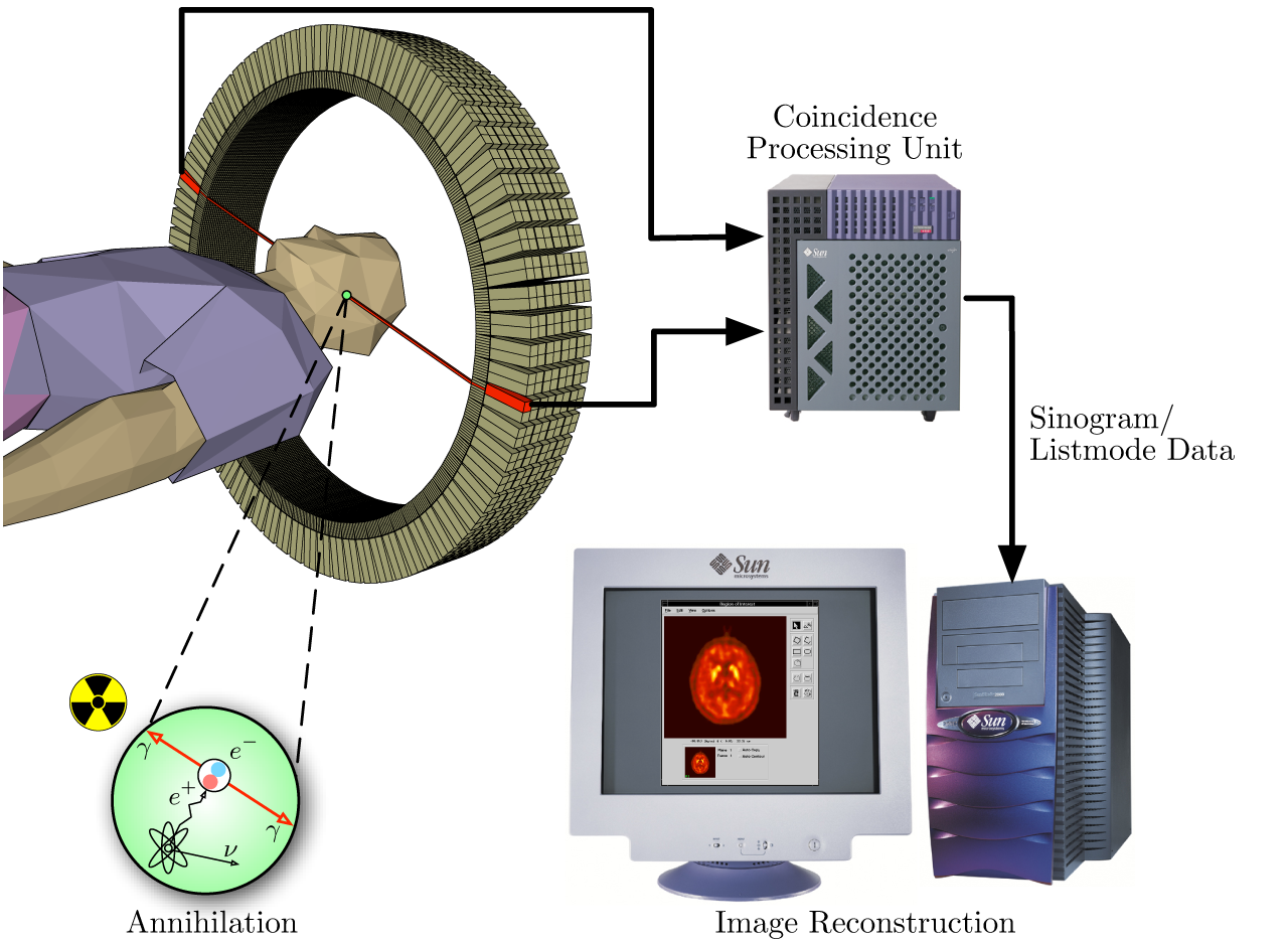
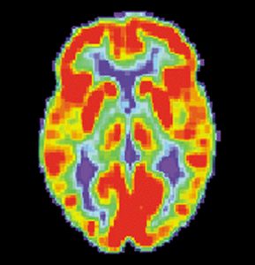
PET is employed both as a medical and research tool. As seen in Figure 1.8, a short-lived radioactive isotope is injected into the participant. The most commonly used is F-18 labeled fluorodeoxyglucose (FDG). After a waiting period for the active molecule to become concentrated in the brain tissue (one hour for FDG), the participant is placed inside the scanner. As the tracer decays, its emissions are collected by the scanner. The scanner depends on detecting a pair of photons moving in opposite directions. Photons that do not have a temporal pair are ignored. Computational reconstruction uses statistical analysis and error correction to produce images such as Figure 1.8 which shows a scan of an unimpaired participant.
As you can imagine, the main issue with PET is the injection of radioactive material into the body. Various jurisdictions set standards on the maximum amount of radiation that a person can be exposed to in a year. This means that the same participant can only take part in a small number of PET scans which limits the amount of data collection possible in psychological studies. Another factor is the expense of PET scanners and the radioactive tracers.
Functional Magnetic Resonance Imaging (fMRI)
An alternative to PET that doesn’t use radioactive substances is Magnetic Resonance Imaging (MRI). This employed powerful electromagnets to affect hydrogen atoms. Hydrogen atoms are abundant in humans as water and fat. The atomic nuclei of hydrogen atoms are able to absorb radio frequency energy when placed in a magnetic field. The resulting spin polarization can produce a radio frequency signal that can be detected and analyzed. Varying the parameters of the radio pulse sequence can produce different contrasts between brain tissues based on the properties of their constituent hydrogen atoms. Computational processing of the signals can produce a highly detailed 3D image of the brain. However, this is a static image of the tissues without any indication of brain activity.
Recently, fMRI (functional magnetic resonance imaging) has come to the forefront as a way to overlap MRI scans with images of brain activity. This measures the energy released by hemoglobin in the blood. It is assumed that the areas of the brain that are most active would be the most likely to take in more blood (for energy). Therefore, the measurement of blood flow with different brain regions can indirectly show us a measure of their activation during particular cognitive tasks. This type of scan provides a better temporal and spatial resolution than PET. However, as there is a 1-5 second lag between brain activation and detection, the temporal resolution of fMRI is inferior to EEG.
Comparing Brain Imaging Technology
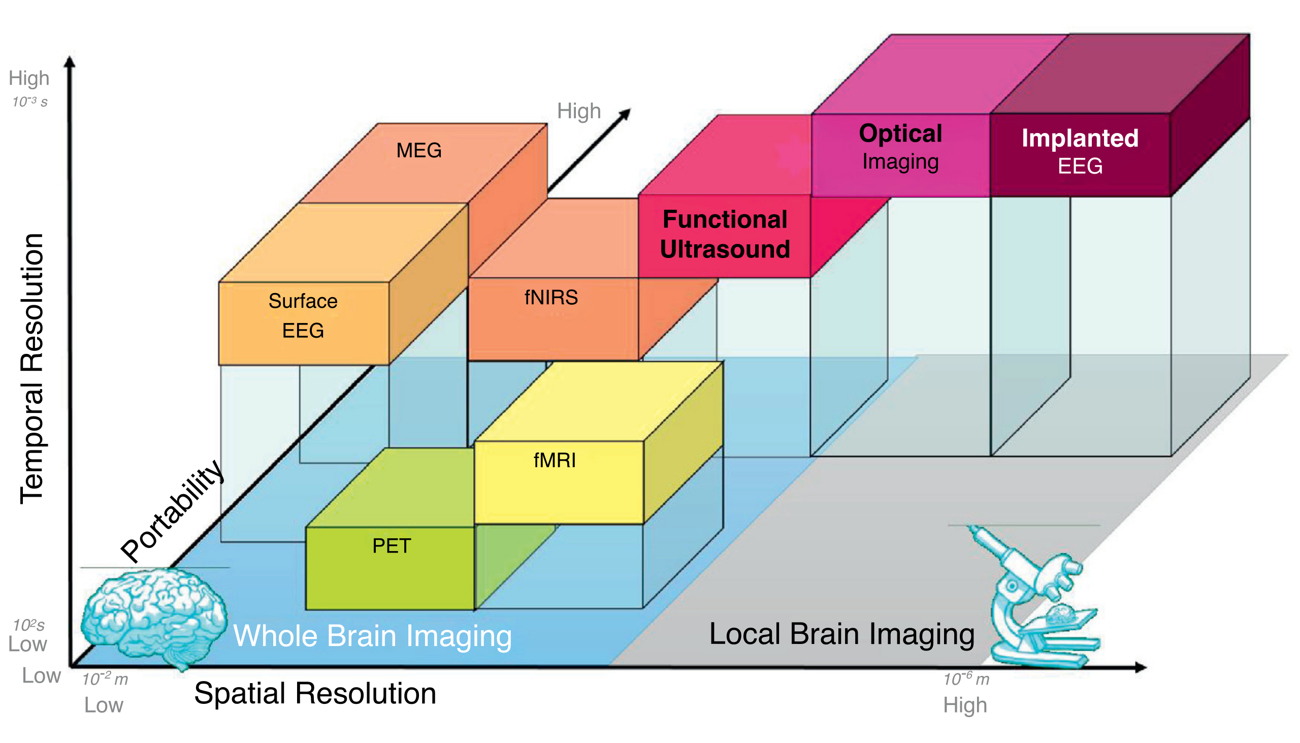
Neuroimaging is at the forefront of psycholinguistic research into language processing in the brain. They can tell us about the time course of various cognitive processes and the extent to which mental processes interact with each other. However, these techniques are still quite expensive and vary in terms of their temporal and spatial resolutions. As can be seen in Figure 1.10, different techniques vary in terms of how accurately they measure timing and active brain regions. EEG can detect brain activity with high temporal resolution but cannot tell us exactly where they originated. As signals are all detected on the surface of the head, we cannot be sure whether they originated in the cortex or areas deeper inside the brain. On the other hand, PET and fMRI are quite good at providing spatial information. However, as they rely on the flow of fluids (blood), there is a temporal lag between when a brain region become active and when the signal is detected by the scanner.
Methodological limitations also exist as most of these techniques require the participant to be still during the scan. This limits the ability to study overt speech or other movement. In addition, the use of powerful magnets in fMRI means that participants with any metal implants cannot take part in such studies (the metal would fly out of their body towards the scanner).
A more serious limitation of any neuroimaging technique is the difficulty in interpreting the results. How do we know what is causing a particular activity? We can see when or where something is happening, but not necessarily how. Observing neural activity is not the same as observing mental activity. Some studies often average out the results from multiple participants. How can we be sure that all of them are using the same brain regions for similar activities? However, even with such limitations, these methods have opened us to a wide range of insights into the neurological basis of language. As new methods are developed, we may even see these methods employed regularly for research and rehabilitation.
Image descriptions
Figure 1.5 Reaction Time Experiment
A diagram showing the process of testing someone’s reaction time to seeing a number on a computer screen and pressing the number on their keyboard:
- Stimuli: The number 3 appears on the computer screen. The timer starts.
- Sensory: The eyes see the number 3.
- Cortical: The stimuli is processed by the brain.
- Motor: The brain tells the hand to press the number 3 on the keyboard.
[Return to the place in text (Figure 1.5)]
Figure 1.7 Positron Emission Tomography Schema
Function of a PET machine. A scanner detects the emissions of the short-lived radioactive isotope in the brain of the subject, transmits this information to a Coincidence Processing Unit, which is subsequently used to reconstruct an image of the subject’s brain activity.
[Return to place in the text (Figure 1.7)]
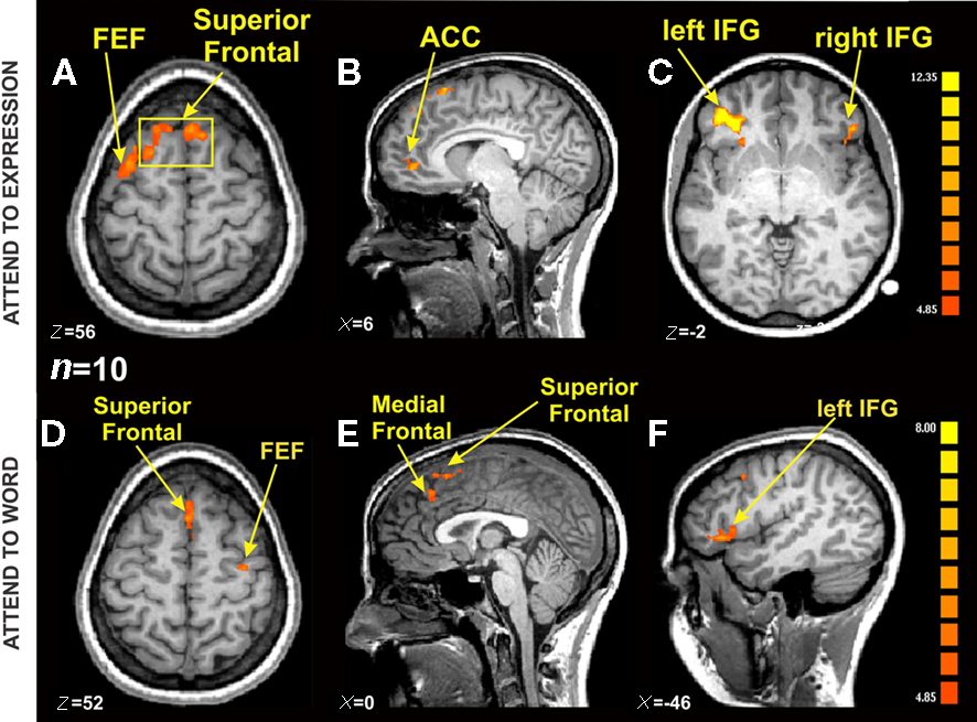
Figure 1.9 fMRI Activation in an Emotional Stroop Task
fMRI scans of six brains. The first three images display the brain’s response to expressions, while the last three illustrate the brain’s response to words. Coloured marks from red to yellow are used to qualitatively assess the strength of the brain’s response, in addition to the location of brain activity.
[Return to place in the text (Figure 1.9)]
Figure 1.10 Comparing Brain Imaging Techniques
A labeled, three-dimensional graph comparing the several brain imaging techniques on the axes of Temporal Resolution, Portability, and Spatial Resolution.
Whole brain imaging techniques listed by spatial resolution from low to high:
- Surface EEG: low spatial resolution, medium portability, high temporal resolution
- MEG: low spatial resolution, high portability, high temporal resolution
- PET: low spatial resolution, low portability, low temporal resolution
- fNIRS: low spatial resolution, high portability, medium temporal resolution
- fMRI: low spatial resolution, low portability, medium temporal resolution
- Functional Ultrasound
Local brain imaging techniques listed by spatial resolution from low to high:
- Optical imaging: high spatial resolution, high portability, high temporal resolution
- Implanted EEG: high spatial resolution, high portability, high temporal resolution
[Return to place in the text (Figure 1.10)]
Media Attributions
- Figure 1.5 Reaction Time Experiment by Emily Willoughby is licensed under a CC BY-SA 4.0 licence.
- Figure 1.6 A Model Priming Web by Noahrob is licensed under a CC BY-SA 4.0 licence.
- Figure 1.7 Positron Emission Tomography Schema by Jens Maus is in the Public Domain.
- Figure 1.8 A PET Scan of an Unimpaired Brain by the US National Institute on Aging, Alzheimer’s Disease Education and Referral Center is in the Public Domain.
- Figure 1.9 fMRI Activation in an Emotional Stroop Task by Shima Ovaysikia, Khalid A. Tahir, Jason L. Chan and Joseph F. X. DeSouza is licensed under a CC BY 2.5 licence.
- Figure 1.10 Comparing Brain Imaging Techniques by Thomas Deffieux, Charlie Demene, Mathieu Pernot, Mickael Tanter is licensed under a CC BY 4.0 licence.
The temporal measure of the time taken between detecting a stimulus and the response to that stimulus.
A phenomenon where exposure to a stimulus influences the response time to a subsequent stimulus.
A damaged or abnormally changed tissue caused by disease or trauma.
Electroencephalography (EEG) is an electrophysiological measurement technique used to record electrical activity on the scalp. This activity represents the electoral activity on the surface of the brain underneath the scalp.
Positron emission tomography is a technique that uses radioactive substances to measure metabolic changes and other physiological changes such as blood flow.
Functional magnetic resonance imaging or functional MRI is a measurement technique used to detect changes in blood flow within the brain.
The way humans use language to communicate and how it is processed and comprehended.
