Central Nervous System Regulation, Mood, and Cognition
8.2 CNS Regulation, Mood, and Cognition Concepts
Concepts Related to the CNS, Mood, and Cognition
This resource provides a basic introduction to the concepts related to the central nervous system and cognition in connection with pharmacology. The concept of cognition is defined as “the process of thought that embodies perception, attention, visuospatial cognition, language, learning, memory, and executive function with the higher-order thinking skills of comprehension, insight, problem-solving, reasoning, decision making, creativity, and metacognition”[1].
This chapter also addresses mood[2], affect[3], and anxiety[4].
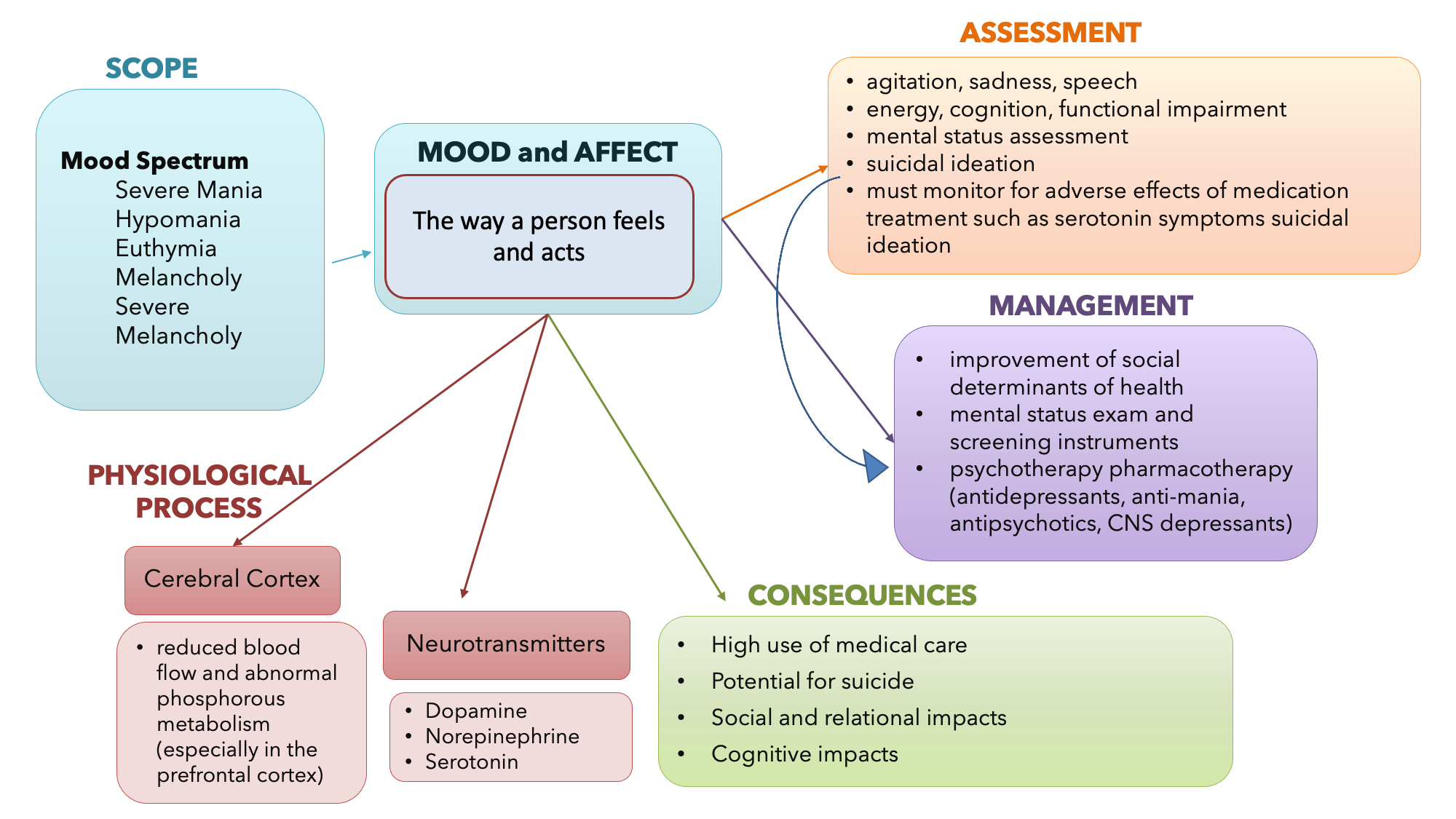
The concept map in Figure 8.2a summarizes information related to the concept of mood, affect, and the CNS [5]. You are encouraged to revisit this map after you have completed the chapter. You may also wish to develop your own concept map related to other CNS concepts in this chapter.
Overview of the Central Nervous System and Processes
Before we can begin to understand how different medications influence the brain, we need to review the central nervous system. The nervous system can be divided into two major regions: the central and peripheral nervous systems. The central nervous system (CNS) is the brain and spinal cord, and the peripheral nervous system (PNS) is everything else. The brain is contained within the cranial cavity of the skull, and the spinal cord is contained within the vertebral cavity of the vertebral column.
It is a bit of an oversimplification to say that the CNS is what is inside these two cavities and the peripheral nervous system is outside of them, but that is one way to start to think about it. In actuality, there are some elements of the peripheral nervous system that are within the cranial or vertebral cavities. The peripheral nervous system is so named because it is on the periphery—meaning beyond the brain and spinal cord. Depending on different aspects of the nervous system, the dividing line between central and peripheral is not necessarily universal.
The peripheral nervous system is further divided into the autonomic nervous system and the somatic nervous system, which are further discussed in the Autonomic Nervous System chapter in this book.[6] (See Figures 8.2b[7] and 8.2c[8] for illustrations of the central and peripheral nervous systems).


Review more detailed information about the nervous system function using this OpenStax link: Basic structure and function of the nervous system
Communication in the Nervous System
Your brain communicates with electrical impulses that signal a release of a neurotransmitter, which then binds to the targeted cell. Understanding this communication will help you put the pieces together when you are trying to understand the mechanism of action of medication that works by influencing neurotransmitters. See Figure 8.2d for an illustration of the major elements in neuron communication.[9]
/10%3A_Overview_of_the_Nervous_System/10.1%3A_Introduction_to_the_Nervous_System/10.1A%3A_Organization_of_the_Nervous_System")
There are two types of connections between electrically active cells: chemical synapses and electrical synapses. In a chemical synapse, a chemical signal—namely, a neurotransmitter—is released from one cell and affects another cell. In comparison, in an electrical synapse, there is a direct connection between the two cells so that ions can pass directly from one cell to the next. In this unit, we will be focusing on the communication of a neurotransmitter in a chemical synapse. Once in the synaptic cleft, the neurotransmitter diffuses the short distance to the postsynaptic membrane and can interact with neurotransmitter receptors. Receptors are specific for the neurotransmitter, and the two fit together like a key and lock. One neurotransmitter binds to its receptor and will not bind to receptors for other neurotransmitters, making the binding a specific chemical event.[10] (See Figure 8.2e for an illustration of a synapse.[11]).

When the neurotransmitter binds to the receptor, the cell membrane of the target neuron changes its electrical state, and a new graded potential begins. If that graded potential is strong enough to reach threshold, the second neuron generates an action potential. The target of this neuron is another neuron in the thalamus of the brain, the part of the CNS that acts as a relay for sensory information. The thalamus then sends the sensory information to the cerebral cortex, the outermost layer of gray matter in the brain, where conscious perception of that stimulus begins.[12]
A supplementary video explaining neuron communication via action potentials is provided below.
Neuron communication via Action Potentials[13]
Types of Neurotransmitters
Amino Acids
One group of neurotransmitters is amino acids. GABA (gamma-aminobutyric acid) is an example of an amino acid neurotransmitter. They each have their own receptors and do not interact with each other. Amino acid neurotransmitters are eliminated from the synapse by re-uptake. A pump in the cell membrane of the presynaptic element, or sometimes a neighbouring glial cell, will clear the amino acid from the synaptic cleft so that it can be recycled, repackaged in vesicles, and released again.
Biogenic Amine
Another class of neurotransmitters is the biogenic amine, a group of neurotransmitters that are enzymatically made from amino acids. For example, serotonin is made from tryptophan. It is the basis of the serotonergic system, which has its own specific receptors. Serotonin is transported back into the presynaptic cell for repackaging.
Other biogenic amines are made from tyrosine and include dopamine, norepinephrine, and epinephrine. Dopamine is part of its own system, the dopaminergic system, which has dopamine receptors. Norepinephrine and epinephrine belong to the adrenergic neurotransmitter system. The two molecules are very similar and bind to the same receptors, which are referred to as alpha- and beta-receptors. The biogenic amines have mixed effects. For example, dopamine receptors that are classified as D1 receptors are excitatory, whereas D2-type receptors are inhibitory.
The important thing to remember about neurotransmitters and signaling chemicals is that the effect is entirely dependent on the receptor.[14]
Functions of Neurotransmitters
An alteration in CNS function is related to abnormal impulse transmission and can result in an imbalance of a neurotransmitter. A person with an imbalance of neurotransmitters may have signs and symptoms of a CNS disorder. The medications that are used to treat CNS disorders mimic or block the neurotransmitter based on the imbalance caused by the condition. Medications are used to either stimulate or depress the effect of the neurotransmitter. For example, CNS depressants alter the brain by decreasing the excitability of neurotransmitters, blocking their receptor site, or increasing the inhibitory neurotransmitter. On the other hand, CNS stimulants increase brain activity by increasing the excitability of neurotransmitters, decreasing the inhibitory neurotransmitters, or blocking their receptor sites.[15]
Norepinephrine is often associated with the fight-or-flight response. Abnormal levels of this neurotransmitter are also associated with depression, decreased alertness and interest, along with possible palpitations, anxiety, and panic attacks. Dopamine is strongly linked to motor and cognition. This neurotransmitter influences movement and can be associated with ADHD, paranoia, and schizophrenia. Serotonin is heavily involved in many bodily processes. Abnormal levels of serotonin can affect sleep, libido, mood, and temperature regulation. Alterations of this neurotransmitter have been linked to many mental health issues such as depression, bipolar disorder, anxiety, and body disorders. GABA (gamma-aminobutyric acid) can act as an inhibitory neurotransmitter. GABA assists with communication in the brain, and if this neurotransmitter is low, it has been linked to issues such as anxiety, seizures, mania, and impulse control. The neurotransmitter glutamate works as an excitatory neurotransmitter and works with GABA to control other functions of the brain.[16]
Image Descriptions
Figure 8.2a Mood and Affect Concept Map
Mood and affect – the way a person feels and acts.
Scope – mood spectrum:
- severe mania
- hypomania
- euthymia
- melancholy
- severe melancholy
Physiological process
- Cerebral cortex
- reduced blood flow and abnormal phosphorous metabolism (especially in the prefrontal cortex)
- Neurotransmitters
- dopamine
- norepinephrine
- serotonin
Assessment
- agitation, sadness, speech
- energy, cognition, functional impairment
- mental status assessment
- suicidal ideation
- must monitor for adverse effects of medication treatment such as serotonin symptoms and suicidal ideation
Management
- improvement of social determinants of health
- mental status exam and screening instruments
- psychotherapy pharmacotherapy (anti-depressants, anti-mania, antipsychotics, CNS depressants).
Consequences
- high use of medical care
- potential for suicide
- social and relational impacts
- cognitive impacts
Figure 8.2c Somatic, Autonomic, and Enteric Structures of the Nervous System
Brain (CNS)
- perception and processing of sensory stimuli (somatic/autonomic)
- execution of voluntary motor responses (somatic)
- regulation of homeostatic mechanisms (autonomic)
Spinal cord (CNS)
- initiation of reflexes from ventral horn (somatic) and lateral horn (autonomic) gray matter
- pathways for sensory and motor functions between periphery and brain (somatic/autonomic)
Nerve (PNS)
- fibers of sensory and motor neurons (somatic/autonomic)
Ganglia (PNS)
- reception of sensory stimuli by dorsal root and cranial ganglia (somatic/autonomic)
- relay of visceral motor responses by autonomic ganglia (autonomic)
Digestive tract (ENS)
- the enteric nervous system (ENS), located in the digestive tract, is responsible for autonomous functions and can operate independently of the brain and spinal cord.
- Jean Giddens, Concepts of Nursing Practice – 2nd edition (Missouri: Elsevier, 2017), page 319. ↵
- Jean Giddens, Concepts of Nursing Practice – 2nd edition (Missouri: Elsevier, 2017), page 299. ↵
- Jean Giddens, Concepts of Nursing Practice – 2nd edition (Missouri: Elsevier, 2017), page 299. ↵
- Jean Giddens, Concepts of Nursing Practice – 2nd edition (Missouri: Elsevier, 2017), page 310. ↵
- Jean Giddens, Concepts of Nursing Practice – 2nd edition (Missouri: Elsevier, 2017) ↵
- This work is a derivative of Anatomy and Physiology by OpenStax licensed under CC BY 4.0. Access for free at https://openstax.org/books/anatomy-and-physiology/pages/1-introduction ↵
- "1201 Overview of Nervous System.jpg" by OpenStax is licensed under CC BY 4.0. Access for free at https://openstax.org/books/anatomy-and-physiology/pages/12-1-basic-structure-and-function-of-the-nervous-system ↵
- "1205 Somatic Autonomic Enteric StructuresN.jpg" by OpenStax is licensed under CC BY 4.0. Access for free at https://openstax.org/books/anatomy-and-physiology/pages/12-1-basic-structure-and-function-of-the-nervous-system ↵
- "Chemical synapse schema cropped.jpg" by Looie496 is licensed under public domain. Access for free at https://med.libretexts.org/Bookshelves/Anatomy_and_Physiology/Book%3A_Anatomy_and_Physiology_(Boundless)/10%3A_Overview_of_the_Nervous_System/10.1%3A_Introduction_to_the_Nervous_System/10.1A%3A_Organization_of_the_Nervous_System ↵
- This work is a derivative of Anatomy and Physiology by OpenStax licensed under CC BY 4.0. Access for free at https://openstax.org/books/anatomy-and-physiology/pages/1-introduction ↵
- "1225 Chemical Synapse.jpg" by Young, KA., Wise, JA., DeSaix, P., Kruse, DH., Poe, B., Johnson, E., Johnson, JE., Korol, O., Betts, JG., & Womble, M. is licensed under CC BY 4.0 Access for free at https://openstax.org/books/anatomy-and-physiology/pages/12-5-communication-between-neurons ↵
- This work is a derivative of Anatomy and Physiology by OpenStax licensed under CC BY 4.0. Access for free at https://openstax.org/books/anatomy-and-physiology/pages/1-introduction ↵
- Forciea, B. (2015, May 12). Anatomy and Physiology: Nervous System: Action Potential Generation V2.0. [Video]. YouTube. All rights reserved. Video used with permission. https://youtu.be/-xFliVq3MKg. ↵
- This work is a derivative of Anatomy and Physiology by OpenStax licensed under CC BY 4.0. Access for free at https://openstax.org/books/anatomy-and-physiology/pages/1-introduction ↵
- This work is a derivative of Pharmacology Notes: Nursing Implications for Clinical Practice by Gloria Velarde licensed under CC BY-NC-SA 4.0. ↵
- This work is a derivative of Pharmacology Notes: Nursing Implications for Clinical Practice by Gloria Velarde licensed under CC BY-NC-SA 4.0. ↵
the process of thought that embodies perception, attention, visuospatial cognition, language, learning, memory, and executive function with the higher order thinking skills of comprehension, insight, problem solving, reasoning, decision making, creativity, and metacognition
the way a person feels [1].
the observable response a person has to his or her own feelings, [2]
an alert to the human condition of impending doom, either real or imagined, and is accompanied by autonomic responses that serve as protective [3].
Anatomical division of the nervous system located within the cranial and vertebral cavities, namely the brain and spinal cord.
An anatomical division of the nervous system that is largely outside the cranial and vertebral cavities, namely all parts except the brain and spinal cord.
Chemical signal that is released from the synaptic end bulb of a neuron to cause a change in the target cell.
Cells that carry electrical impulses to the synapse of a target organ.
Connection between two neurons, or between a neuron and its target, where a neurotransmitter diffuses across a very short distance.
Connection between two neurons, or any two electrically active cells, where ions flow directly through channels spanning their adjacent cell membranes.
The membrane voltage at which an action potential is initiated.
A change in voltage of a cell membrane in response to a stimulus that results in transmission of an electrical signal; unique to neurons and muscle fibers.
The region of the central nervous system that acts as a relay for sensory pathways.
